
Sarcomas - malignant tumors in soft tissue or in the bones
Sarcomas can develop almost anywhere in the body and their symptoms are difficult to recognize because they often cause little pain and are often dismissed as harmless swellings by those who suffer from them. Because these tumors are often detected very late at an advanced stage and treated by non-specialists, the prognosis is worse compared to other types of tumors. The most common bone sarcomas are osteosarcoma, chondrosarcoma, and Ewing's sarcoma. Soft tissue sarcomas are more common and can occur in any soft tissue such as fat tissue, muscle tissue, or nerve tissue. Sarcomas should be treated in specialized sarcoma centers with appropriate experience and expertise. Due to the relative rarity of bone and soft tissue sarcomas, this type of cancer remains one of the most difficult to treat.
The specialists from Heidelberg University Hospital
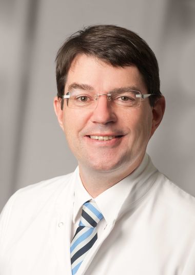
Prof. Burkhard Lehner, MD, is Head of the Section for Oncological Orthopedics and Septic Orthopedic Surgery and former Medical Director of the Clinic for Orthopedics and Trauma Surgery of the Sarcoma Center at Heidelberg University Hospital. He specializes in bone cancer and soft tissue sarcomas, particularly osteosarcomas, Ewing sarcomas, chondrosarcomas, bone metastases, giant cell tumors, and soft tissue tumors. Professor Lehner has special expertise in prosthetics and biological reconstruction of bone defects.
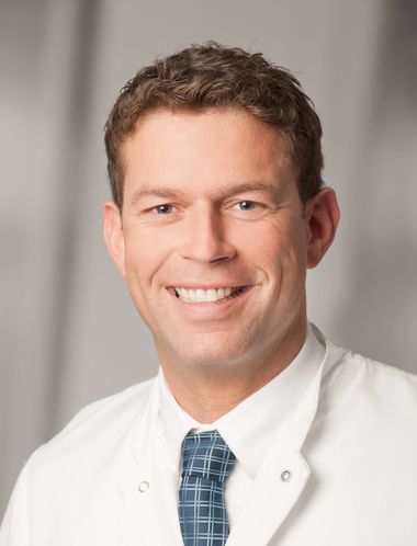
Stefan Hemmer, MD, is the head of the spine surgery ward at the Sarcoma Center at Heidelberg University Hospital. He specializes in the evaluation of the stability of spinal tumors, resection of tumors and reconstruction of the spine, treatment of scoliosis and spinal deformities, complex treatment of pediatric spinal diseases, treatment of spinal stenosis and all types of spinal fractures.
Frequently Asked Questions (FAQ)
The specialists from Heidelberg University Hospital, Stefan Hemmer and Burkhard Lehner, provide information on the initial symptoms of sarcomas and on modern approaches to prosthetics and rehabilitation.
What causes a sarcoma?
There is no definite cause for sarcoma that can be named. Genetics plays a rather minor role. Radiation therapy in conjunction with another disease can, in rare cases, contribute to the development of sarcoma. Environmental toxins and chemicals such as wood preservatives, herbicides, dioxin or arsenic as well as chronic inflammation also seem to promote the formation of soft tissue sarcomas.
Why sarcoma is difficult to treat?
Performing surgery to treat a sarcoma requires a high level of experience. During an operation, the tumor must be removed completely in healthy tissue while preserving the extremity and the function must be affected as little as possible. The surgeon has to master all possibilities of tumor resection and also of reconstruction of a bone defect after bone tumor resection. Appropriate special tumor implants must be available. In addition, an individual treatment plan based on the patient's exact histology must be drawn up in a specialized center, which also takes into account possible additional forms of therapy such as chemotherapy or radiotherapy.
Since sarcomas are uncommon cancers, the awareness of it stays low. What groups of the population are more likely to get sarcoma?
Men are more likely to be affected than women. Soft tissue sarcomas are rare with an incidence of 2-3 new cases per 100,000 inhabitants per year and account for 1-2% of all malignant tumors in adults. In Germany, about 3000 new cases per year are expected. In children, soft tissue sarcomas have a significantly higher proportion of malignant tumors (about 5%). Soft tissue sarcomas can occur at any age, but are more common between the ages of 45 and 55 or under the age of 15.
What are the first symptoms in the early stages?
There are no characteristic symptoms for sarcomas. This is why sarcomas are often detected late. Most sarcomas initially cause a painless swelling or lump. As they grow, a feeling of tightness may develop in the affected area. If the sarcoma pushes out nerves or grows into nerve tissue, pain occurs. Bone sarcomas may cause joint pain, joint stiffness, or a broken bone from the tumor.
What should a patient do, if he thinks he has a sarcoma?
In case of a suspected sarcoma it is especially important to find competent specialists who have a lot of experience with the diagnosis and treatment of this particular type of tumor.
What tests are done to diagnose sarcoma?
Magnetic resonance imaging (MRI), X-ray, Computed tomography (CT), Bone scintigraphy and Positron emission tomography PET. After imaging, a biopsy is required for histological confirmation. If possible, this biopsy should be performed at the center where the tumor is then operated on. If the biopsy is performed in the wrong way, it can significantly worsen the patient's prognosis.
If the diagnosis is confirmed, how should one choose a clinic for treatment? What kind of facilities can characterize a proper center for curing sarcomas?
It is always best for patients to consult a large center that has the greatest possible experience. Sometimes more can be done for sufferers here or experienced experts confirm the therapy and this gives greater certainty that the right thing is being done. It is also recommended to get a second opinion. Heidelberg University Hospital offers medical second opinions and video consultations for this purpose. At the National Center for Tumor Diseases, bone and soft tissue sarcomas are diagnosed and treated on an interdisciplinary basis. The Heidelberg Sarcoma Center is characterized by the excellent interdisciplinary cooperation of the different specialties. In the weekly sarcoma boards, approximately 600 patient cases are discussed and treatments are determined each year. Orthopedic surgeons, visceral surgeons, plastic surgeons, medical oncologists, radiation therapists, radiologists, pathologists, and representatives of other departments participate in this sarcoma board to determine the appropriate treatment strategy for each individual case of disease. Most sarcomas occur in the extremities and spine and can only be treated sufficiently by surgical removal by the orthopedic surgeons at Heidelberg University Hospital.
If treating sarcoma requires surgery, what is supposed to happen afterwards?
After a successful operation, an individual rehabilitation plan is developed to restore the function of the operated limb or back as much as possible. Physiotherapy treatment is already provided during the inpatient stay for the operation in order to mobilize the patient again as quickly as possible. In addition, an individual post-surgical care plan (for a period of five (5) to ten (10) years) is provided. Initially, follow-up appointments may take place every three months, but the examination intervals may be extended in the future. The follow-up appointments can be performed on an outpatient basis, or in a specialist's office or in specialized clinics. Particular attention is paid to whether new tumor cells are formed and then treat them quickly.
For a while, the most common way to treat sarcoma was amputation, however, it seems that now a number of limb-sparing techniques can be available for patience as well. Could you tell about them?
The Sarcoma Center in Heidelberg with its highly qualified specialists uses modern implants made of titanium. In spinal orthopedics, implants made of carbon are also used so that subsequent diagnostics in MRI and CT can continue to be max. efficient and meaningful. This can also or a possible postoperative radiation therapy with heavy ions be made possible. In addition, silver-coated implants to prevent infection or patient-specific implants (3D manufactured from titanium with a special surface to optimize bony ingrowth) are used for extremities and pelvis. For the reconstruction of large bone defects, especially in the joint area, the most modern tumor endoprostheses with silver coating or custom-made metal implants are used. If possible, reconstruction is carried out biologically using the patient's own graft bone or foreign bone.
Are there any new approaches in treating sarcoma using chemotherapy, radiation therapy (also called "radiotherapy") and proton therapy? Any innovations in order to reduce side effects?
For most malignant bone tumors and for many highly malignant soft tissue tumors, individually adjusted chemotherapy is performed in addition to surgery either before or after surgery. Radiation therapy may be required for some bone tumors. Highly malignant soft tissue tumors are usually additionally irradiated either before or after surgery. If possible, in the form of proton irradiation or heavy ion irradiation. In some cases, ion beams are the best possible radiation therapy, as they are particularly effective, can be controlled with high precision, and cause hardly any side effects. Proton therapy and heavy ion therapy hit the tumor much more precisely and deliver their therapeutic energy accurately in the tumor - surrounding healthy tissue is spared and there are fewer side effects. Gene typing is performed for all sarcomas at the Heidelberg Sarcoma Center to enable individualized therapy. In the future, highly precise forms of treatment with chemotherapy or immunotherapy can be individually adapted to each tumor.
Can a sarcoma come back? What factors can determine the perspectives of patient?
Sarcoma recurrences can occur as with all tumor diseases. These can occur locally as a local recurrence or in the form of metastases, usually in the lungs. In order to detect recurrence or metastasis at an early stage, regular follow-up is performed by means of examination and MRI of the operated area and a CT scan of the lung. If possible, surgical removal of these local recurrences or metastases is performed, as this is associated with a better prognosis. Systemic therapy will often be required (chemotherapy or targeted therapy, participation in a clinical trial if necessary).
Further information
Heidelberg University Hospital & Faculty of Medicine
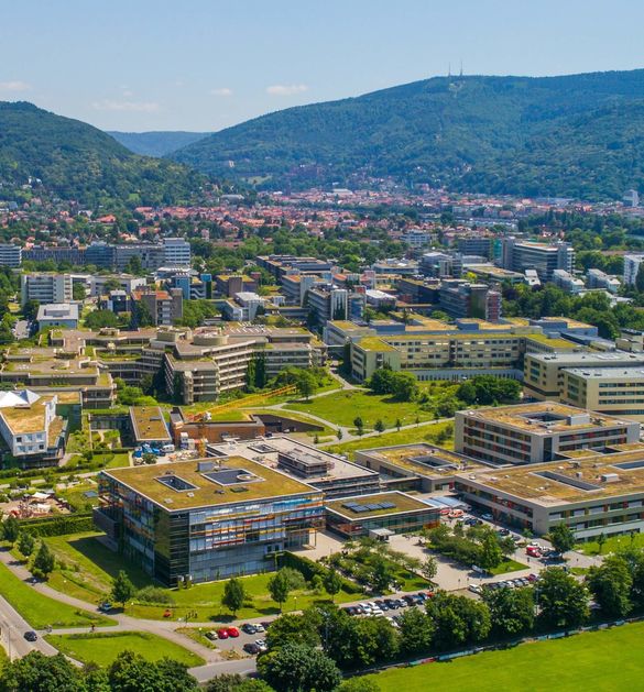
The Heidelberg University Hospital and Faculty of Medicine: internationally are renowned for Patient care, research and teaching. Heidelberg University Hospital is one of the most important medical centers in Germany; the Heidelberg Medical Faculty of Heidelberg University is one of the internationally renowned biomedical research institutions in Europe. The common goal is the development of innovative diagnostics and therapies and their rapid implementation for patients. The hospital and faculty employ around 13,700 staff and are committed to training and qualification. In more than 50 clinical departments with almost 2,000 beds, approximately 80,000 patients are treated annually as full and partial inpatients and more than 1,000,000 patients are treated as outpatients. Together with the German Cancer Research Center (DKFZ) and German Cancer Aid, Heidelberg University Hospital has established the National Center for Tumor Diseases (NCT) in Heidelberg, which is the leading oncological center of excellence in Germany. In addition, Heidelberg University Hospital, together with the DKFZ and Heidelberg University, operates the Hopp Children's Tumor Center (KiTZ), a therapy and research center for oncological and hematological diseases in childhood and adolescence that is unique in Germany. The Heidelberg Curriculum Medicinale (HeiCuMed) is one of the top medical training programs in Germany. There are currently around 3,500 future physicians studying for their degrees and doctorates at the Heidelberg Medical Faculty.
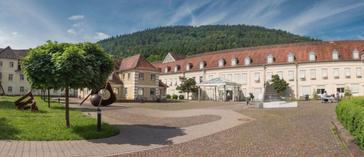
The Sarcoma Center in Heidelberg
The Sarcoma Center in Heidelberg with its highly qualified specialists uses modern implants made of titanium. In spinal orthopedics, implants made of carbon are also used so that subsequent diagnostics in MRI and CT can continue to be max. efficient and meaningful. This can also or a possible postoperative radiation therapy with heavy ions be made possible. In addition, silver-coated implants to prevent infection or patient-specific implants (3D manufactured from titanium with a special surface to optimize bony ingrowth) are used for extremities and pelvis. For the reconstruction of large bone defects, especially in the joint area, the most modern tumor endoprostheses with silver coating or custom-made metal implants are used. If possible, reconstruction is carried out biologically using the patient's own graft bone or foreign bone.
Orthopedic Oncology
For over 20 years, we have been providing the highest level of care for patients with tumors of the bone and soft tissue in the Department of Orthopedics and Traumatology. Our highly experienced, specialized team is almost exclusively engaged in the comprehensive diagnosis and effective treatment of benign and malignant tumors of the bone and soft tissues.
Our areas of concentration within the field of orthopedic oncology are based on procedures and management concepts that preserve extremities as well as posture and mobility, for example, with the help of special bone and soft tissue transplants.
In collaboration with other departments we employ the most modern therapeutic measures: neo-adjuvant chemotherapy, intraoperative radiation therapy, and extracorporeal irradiation of bone tumors with the goal of preserving the extremity. Also reconstruction of bone defects in the region of joints with a modular tumor endoprosthesis system is a common procedure in our department.
We specialize in treating
- Osteosarcoma
- Ewing sarcoma
- Chondrosarcoma
- Bone metastases
- Giant cell tumors
- All kinds of benign bone tumors
- Soft tissue sarcomas
Spinal Surgery
At the Center for Spinal Surgery, we provide care to approximately 3300 ambulatory and 650 in-hospital patients per year. In addition to the most modern non-operative therapies for degenerative diseases of the spine, we offer expertise over the entire operative spectrum of spinal surgery. This includes surgical treatment of the entire spine (cervical, thoracic, and lumbar) at the highest academic level and according to the latest scientific standards.
All surgical techniques, anterior and posterior, are well established in our center. This also applies to more recent minimally invasive surgical techniques such as percutaneous spinal fusion and minimally invasive vertebral body replacement. Intraoperative neuromonitoring is routinely used for all complex spinal deformities. In selected cases, navigation technology is used.
In cooperation with the Department of Radiology, we offer our patients the full range of diagnostic procedures, providing accurate diagnosis prior to surgery so that targeted and efficient treatment can be performed. Our clinic is one of the few hospitals in Germany boasting a low-dose X-ray machine (EOS® imaging with 3D reconstruction), which provides highly informative radiography with very low radiation exposure. This technique is complemented by 128-slice spiral CT and 3 Tesla MRI, plus the availability of CT myelography. In cooperation with the Spinal Cord Injury Center (Neurology), electrophysiological and neuro-urological examination facilities are always available to our patients.
We specialize in treating
- Congenital and acquired diseases of the spine
- Pediatric spinal disorders
- Degenerative diseases of the spine
- Spinal deformities
- Idiopathic scoliosis / neurogenic and neuromuscular scoliosis
- Congenital scoliosis
- Reconstructive spinal surgery
- Spinal fractures
- Infections of the spine
- Surgical spinal oncology
- En Bloc spondylectomy
- Disc surgery
Individual treatment offer
To check if a treatment option / appointment is possible, submit your data here for our medical specialists.
Second opinion service
For international patients the specialists at Heidelberg University Hospital offer the possibility of a second opinion in the form of a written second opinion or a video consultation. This allows to discuss the specific case and possible next steps. After uploading findings via the telemedicine portal, specialists analyze these documents: Findings that have already been collected are reassessed and supplemented if necessary. Patients are supported with this assessment in making a sensible and good decision for further treatment.