
Coiling
Therapy of brain aneurysms
There are basically two different therapy options for aneurysms in the brain: the original method involves opening the cranium and subsequently placing of a metal clip (surgical clipping) across the aneurysm from the outside, and inserting ultrafine platinum coils (endovascular coiling), without a craniotomy, to occlude the aneurysm from the inside. Pursuant to the results of the ISAT study (International Subarachnoid Aneurysm Trail, 2002) and numerous case series, the recommendation for treatment of aneurysms that according to the technical criteria are eligible for coiling is preferably an endovascular approach. Nonetheless the choice of procedure depends on various different factors that need to be discussed with the patient in detail. The Department of Neuroradiology at the Heidelberg University Hospital is engaged in intensive cooperation in this area with the Clinic for Neurology and the Clinic for Neurosurgery, operating as an interdisciplinary team to establish the best possible therapy for each individual patient.
Coiling procedure
Before starting the actual coiling procedure, the patient receives a general anaesthetic to prevent any movement of the head. To reach the aneurysm, the femoral artery in the groin is tapped first and a small tube of approximately 2-4mm (sheath) is inserted. A catheter is then placed through the sheath and positioned in the corresponding carotid artery under fluoroscopic imaging guidance. Then, a three-dimensional angiography is performed which helps to plan the procedure and localize the working projection. A second, very fine catheter (micro-catheter, the diameter of the tip is approx. 0.5mm) is inserted through the catheter and navigated into the aneurysm through the femoral artery in the groin. Platinum coils (diameter approx. 0.25mm), which fill the aneurysm in a spiral, thus gradually fill the aneurysm dome up to its closure. After completion of the coiling, the catheter and sheath are removed, and the groin is closed with a fibrin clot. Subsequently, the patient awakens in the intensive care unit and typically remains there one night for monitoring.
Remodeling and Stent Implantation
Depending on the size, position and direction of the aneurysm, the Heidelberg University Hospital regularly uses vessel-reconstructing methods such as the so-called “remodeling” technique with balloon-assisted aneurysm filling or insertion of stents. By using these supplementary measures, it is possible today to treat the majority of intracranial aneurysms through an endovascular approach by coiling.
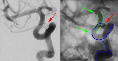
The red arrow in both illustrations shows an aneurysm (weak spot in vascular wall) of the right front cerebral artery (A. carotis interna right, front view). For the safe treatment of this aneurysm, a tubular metal mesh (stent) was first inserted in the vascular wall and precisely placed above the mouth of the aneurysm. The almost invisible stent is marked with green dotted lines on the right part of the illustration. On both of its ends, it is marked with visible dark markers which the green arrows are pointing out. The purpose of a stent in the treatment of aneurysms is to narrow the mouth of the aneurysm with a mesh. Then, the platinum coils can be inserted safely in the aneurysm through the mesh. The platinum coils seal the aneuryms and can, in difficult cases, be better held in the aneurysm due to the underlying stent. The platinum coils are inserted in the aneurysm via a very fine, tiny plastic tube (so-called micro-catheter) through the mesh. In this case, the path of the micro-catheter is drawn with a blue dotted line.