We treat (Adults and Children):
- Primary brain tumors, e.g. pituitary tumors, gliomas, astrocytoma
- Brain metastases
- Basal skull tumors, e.g. clivus chordoma, basal skull meningioma
- Tumors of the meninges, meningioma
- Spinal cord tumors
- Tumors of the spine, e.g. chordoma, chondrosarcoma
- Brain and spinal cord tumors in children, e.g. ependymomas, medulloblastoma
State-of-the-art operative techniques
...are applied daily in our five operating theaters and individualized for patients
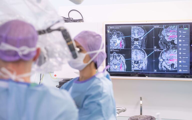
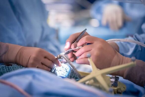
- Computer-assisted surgical instruments (Neuronavigation systems) and modern surgical microscopes (microsurgery) are standard: Thus, the tumor can be reached and eliminated to the millimeter.
- Staining of tumor tissue with fluorescent substances: The neurosurgeon can distinguish the tumor from healthy tissue
- Endoscopic procedures: Without opening the skull, e.g. procedures through the nose
- Awake craniotomy: For tumors in the language center, the procedure may be performed with the patient intermittently awake (pain-free), to monitor and maintain language function during tumor removal. Patients are carefully prepared for the operation by trained psychologists and cared for during the procedure.
Treatment by experienced experts and a fixed physician as contact person
On request, Prof. Krieg or one of his most experienced deputies who specialize in the particular disease can perform the surgery for International Patients. All patients receive extensive information at the personal consultation and can ask any and all questions they have regarding treatment. Your surgeon is also available after surgery and during follow-up as medical consultant.
Neurosurgical specialist team for children with brain tumors
Specialized neurosurgeons are available for the treatment of children with tumors of the brain or spinal cord. The treatment is always performed in cooperation with the nearby Children's Hospital, and particularly with the Department of Pediatric Oncology / Section for Pediatric Brain Tumors.
For all medical specialties, the following applies:
Compassionate care for our adult and pediatric patients, who are often in a very challenging situation, is equally important to us. Our patients have confirmed our success in this over and over, and this reinforces our approach.
Unique technical diagnostic equipment before and during the operation
- MRI (devices up to 3 Tesla)/CT diagnostics for operative preparation, including visualization of brain functions in the surrounding tumor tissue (Department of Neuroradiology)
- Intraoperative MRI (1.5 Tesla)/CT: Imaging is used during the operation to check whether the tumor has been completely removed or whether further surgery is needed.
- "Buzz on wall": Display of all images within one image and thus, greatest support for the surgeon and safety for the patient.
- Neuromonitoring: During the operation, brain and nerve functions are continuously monitored for rapid detection and minimization of potential damage.
After surgery - what's next?
We'll discuss with you whether further treatment is necessary. Essential decisions are all reached by an interdisciplinary expert team. In our weekly tumor boards, each patient and his/her disease is discussed individually and the optimal treatment plan developed.
The neurosurgeons standing by your side...
- Department of Neuroradiology
- Department of Neuropathology - Tissue diagnostics at the highest international level
- For the treatment of frontal and lateral skull base tumors / clivus chordoma, we operate together with our ENT and Oral and Maxillofacial Surgery colleagues.
- In children, treatment is always performed with a specialized team of pediatric neurosurgeons coordinated with a specialized brain and spinal cord tumor team from the Children's Hospital.
- Department of Neurooncology
- Department of Pediatric Oncology / Section: Pediatric Brain Tumors
- Department of Radiooncology (cyberknife, IMRI, MR-Linac, etc.)
- Heidelberg Ion Beam Therapy Center for Proton / Heavy Ion Radiation (e.g. as alternative therapy for non-operable skull base tumors)
- National Center for Tumor Diseases
-
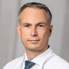
For me and my team, the ultimate goal is to maximize the precision of complete removal of tumor tissue while preserving the surrounding tissue. This is achieved through extensive experience gained from many thousands of operations, a highly-specialized interdisciplinary medical team, the use and development of innovative treatment techniques, state-of-the-art Equipment as well as intensive clinical research activities.
Want to learn if we offer treatment in your case?
How to arrange an appointment.
Please send us, via our international telemedicine portal:
- The completed contact form with important patient information
- A current medical report (in German, English, or Russian)
- MRI / CT images (these should not be older than 3 months)
- The written report of the radiologist from the native country
Once the information file is received in completion, the International Office will direct the request to an experienced neurosurgical expert. We will review the file within several days and inform you whether we can offer treatment at Heidelberg University Hospital Department of Neurosurgery.


