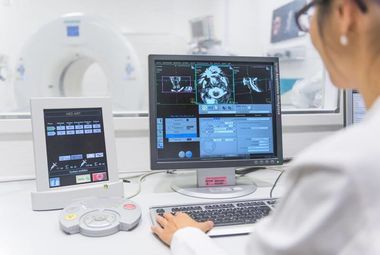
Radiation Dose During Computed Tomography Reduced by Half

November 2017
The Department for Diagnostic and Interventional Radiology at Heidelberg University Hospital was distinguished for outstanding commitment in the area of radiation protection during computed tomography. In Heidelberg, radiation levels have remained markedly below the nationally stipulated levels of Germany and many additional safety precautions are taken that exceed legal requirements. In addition, the hospital has a special head protection that protects the sensitive optic lens from scattered radiation during head CT.
The Department for Diagnostic and Interventional Radiology at Heidelberg University Hospital received five stars by the "EuroSafe Imaging" initiative of the European Society of Radiology. Radiological hospitals and departments Europe-wide are awarded the maximum number of five "EuroSafe Imaging Stars" for implementing the best possible radiation protection measures, continuously researching further improvements, and carrying out the rigorous and extensive certification criteria of the initiative. Thanks to continuous optimization of examination protocols and the use of the latest technology, the Heidelberg Radiology team, headed by Professor Dr. Hans-Ulrich Kauczor, has succeeded in the past several years at reducing average radiation exposure levels by half during computed tomography (CT), and at keeping them well under the legal limits of Germany. Along with Heidelberg, only one single German hospital has been certified with five stars.
With computed tomography (CT), patients are exposed to about ten times the radiation of a normal x-ray. To keep the exposure levels as low as possible, radiologists must master a balancing act between image quality and radiation exposure. The great challenge of diagnostic radiology is to determine a reasonable radiation exposure for each patient and each situation individually, depending on the required image accuracy.
CT System with "Anatomic Understanding"
A new procedure, currently under refinement by several studies, is the so-called iterative reconstruction. The idea is that the CT system gains anatomical understanding through the use of special software. Thus, it "expects" particular structures in particular places - for example, intestinal loops, and sections of the liver, aorta, and spine on a cross-section of the abdomen - and compares the actual image of the patient with this model.
Currently, the procedure is still being adjusted to various investigation scenarios, e.g. for the detection of tumors or for intensive care patients with lines and tubes. There is still room to take the radiation dose of CT even lower. "I assume that with all these new methods that we've introduced to our clinic in the past one and a half years that we can reduce the radiation exposure again by 25 percent," says Kauczor, whose team performs more than 30,000 CT examinations annually.
Further information:
All departments of Heidelberg University Hospital

