
Digital OR in facial surgery
February 2016
Oral and maxillofacial surgery at Heidelberg University Hospital involves a digital OP management system, which enables a more precise operation.
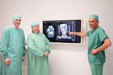
Especially in the field of facial surgery, physicians work with huge quantities of data in order to reconstruct the patient’s original condition following an accident, for example. Now these large quantities of data are bundled and made accessible to surgeons in user-friendly ways with the aid of a digital OR management system. Therefore, they now have the possibility of downloading and viewing the patient’s images and video data during an operation. This allows for a more precise course of the intervention, because the surgeons work with layer images comprising up to 500 individual images. Via large multi-touch screens, the surgeon can access these X-ray images directly from the operating table. Wolfgang Marguerre, a Heidelberg entrepreneur and CEO of Octapharma AG, financed this digital OR Management System with a donation of EUR 250,000. "We are very grateful to Octapharma and Mr. Marguerre for their support, especially on behalf of our patients“, says Professor Dr. Dr. Jürgen Hoffmann, Medical Director of the Department of Oral and Maxillofacial Surgery and Polyclinic at Heidelberg University Hospital.
Virtual models of the face provide support to surgeons during the operation
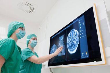
This new technology also allows the documentation of operations. Images or videos, respectively, are recorded through the microscope, endoscope or the camera in the OR lamp. This new system primarily enables complex reconstructions in interventions related to tumors and accidents. “With tumor patients, for example, we first have to remove the lower jaw and then reconstruct it with the body’s own tissue. On the display, which looks like a mega-pad, we have the images of the patient’s skull shown to us three dimensionally and move it all directions”, explains Professor Hoffmann. In the form of a virtual model of the face created from the patient’s images, the surgeons can see the finished results in advance.
International experts are enthusiastic about new conference systems
In advanced and ongoing training, physicians and students benefit from the digital OR management system. They become familiar with the techniques directly in the operating room, or the intervention is broadcast to the neighboring conference room via a video link. Recently, a course in surgery was offered to an international audience at which the new conference system was successfully used. A reconstruction of the lower jaw by means of a bone transplant was performed. Operating surgeons connected the blood vessels of the transplant under the microscope. Surgeons wore headsets in order to explain the procedures to the watching participants. Due to the high-resolution images, physicians were able to follow the operation very well and proved to be enthusiastic about the transmission.
Further information:
Department of Oral and Maxillofacial Surgery